sono tenderness at mcburneys point (or focal appendix tenderness with probe pressure)
thickened appendix: > 6mm transverse diameter
loss of wall stratification ie loss of the ‘target appearrance)
free fluid around the appendix
important: depending on the degree of inflammation and timing of the scan since onset of symptoms, as to how many of these signs may be present. a hyperaemic aptender appendix should be considered as suspicios even without other sono-signs.
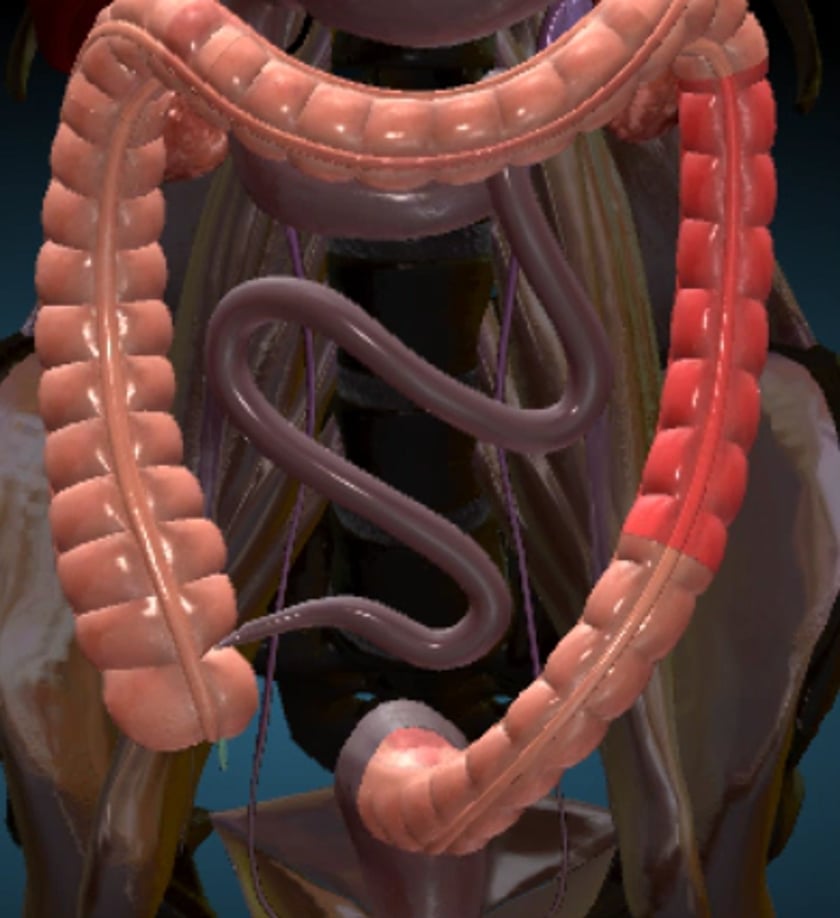
anatomy
the appendix (blue) is seen lying behind the caecum in most cases.
anatomy appendix
appendix retrocaecal position
normal appendix positions
the appendix can be found from the groin, to the umbilicus or even higher under the liver.
the appendix position variations are usually right sided.
it is rarely midline or in the left iliac fossa.
mcburneys point: 1/3 the distance from asis to umbilicus.
refer to this study which concluded that the location of the appendix has wide individual variability,and the limitations of mcburney’s point as an anatomic landmark should be recognised.
visualisation of the appendix in its entirety is required because appendicitis may only affect the tip.
normal appendix- longitudinal axis
appendicitis should be suspected when the outer diameter of the appendix measures greater than 6mm.
scan protocol
role of ultrasound
limitations
bowel gas and patient habitus are the biggest limiting factors to visualising the appendix.
up to 60% of appendices are retrocaecal and thus may be obscured. not identifying an appendix does not exclude appendicitis.
patient preparation
ideally the patient has fasted for 6 hrs. water in the bladder is an advantage to rule out ovarian pathology. unfortunately the appendix is usually an urgent “fit in” and the preparation cannot always be adhered to.
equipment setup
use of a high resolution probe (7-15mhz) is essential. beam steering or compounding can help to overcome anisotropy in linear structures such as tendons. good colour / power / doppler capabilities. be prepared to change frequency output of probe (or probes) to adequately assess both superficial and deeper structures.
common pathology
appendiciitis
mesenteric adenitis
ovarian pathology
crohn’s disease
diverticulitis
abscess
scanning technique
finding the appendix is highly sonographer dependent. they must have a good skill level to undertake this examination.
begin by placing the transducer in a transverse position and applying deep graded compression to the displace the gas and bring the bowel closer to the probe.
beginning at the hepatic flexure the bowel is traced down to the caecum.
the patient should point to the location of pain .
it is a good idea to have a protocol which includes the entire pelvis of all females with right lower quadrant pain and scanning the renal and biliary systems of all patients with a normal appendix.
sometimes the external iliac artery and vein can provide a good landmark for finding the appendix because of the location and pulsatility, compressible, and having doppler flow.
ultrasound criteria to diagnose appendicitis
in order to demonstrate all the possible presentations of appendicitis it is important that the entire appendix is visualized
when the outer diameter of the appendix measures greater than 6 mm
echogenic inflammatory periappendiceal fat change
the wall thickness can measure almost 3 mm or greater
progressed appendicitis can demonstrate a gangrenous appendix. the lumen distends tremendously sometime upwards to 2 cm and is not compressible. an appendicolith may be present which will cast an acoustic shadow.
an appendicolith may be present which will cast an acoustic shadow
or a perforated appendix is demonstrated when the appendicular wall has ruptured producing fluid or a newly formed abscess. the appearance is hyperechoic with an echo-poor abscess surrounding the appendix. there may be a reflective omentum around the appendix, a thickened bowel, and enlarged lymph nodes. asymmetrical wall thickening may indicate perforation.
free fluid in the periappendiceal region
clinical criteria to diagnose appendicitis
there have been numerous signs and suggestions over the preceding 150years.