hand-finger normal

for images of pathology, and detailed descriptions.

palmar aspect

the primary structures to examine are:

  • flexor tendons and sheaths.
  • joints
  • pulleys
  • volar plates
  • neuro-vascular bundles

flexor tendons

thumb:

  • pulleys: 2 annular pulleys and an oblique pulley (which arises from the aponeurosis of the adductor pollicis muscle (thenar eminence)
  • tendons: flexor pollicis longus(fpl) runs through the carpal tunnel and thenar eminence to insert on the distal phalanx. flexor pollicis brevis forms the thenar eminence of the hand.
thumb flexor anatomy labelled
thumb flexor anatomy labelled. ref www.anatomylearning.com

fingers:

  • tendons: there are 2 primary flexor tendons of the 4 fingers:
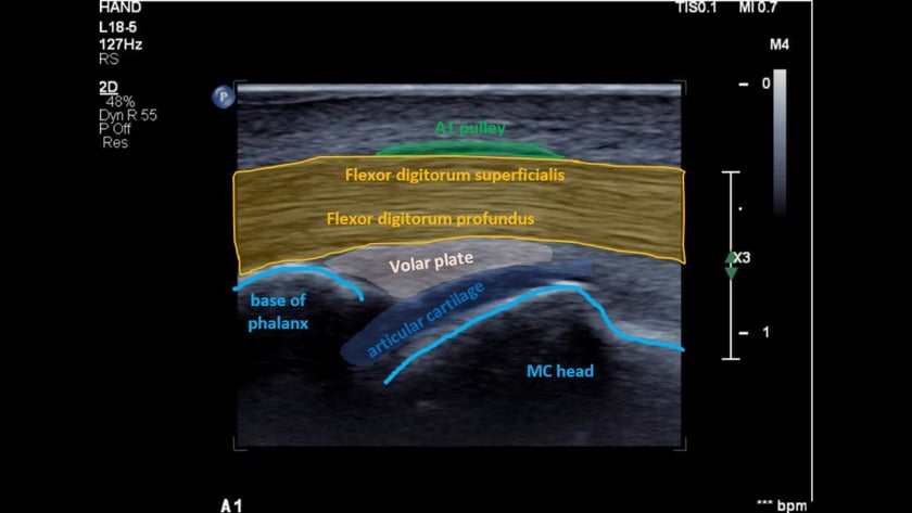
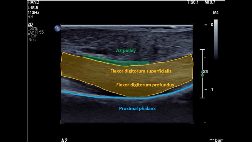
  1. flexor digitorum superficialis tendon(fds). this lies most superficial at the level of the metacarpal. it divides into 2 slips, each circling under the fdp tendon to insert as 2 separate slips onto the base of the middle phalanx. (function: flex the pipj)
  2. flexor digitorum profundus tendon (fdp) is deep to the fds. the fdp inserts onto the base of the distal phalanx. (function: flex the dipj)
  • pulleys: role is to support and guide  the flexor tendons along the bone.

5 annular pulleys (a1-5) and 3 cruciate pulleys (c1-3). the cruciate pulleys are poorly seen on ultrasound due to anisotrophy.

anatomy of the flexor tendons of the fingers labelled
anatomy of the finger pulleys labelled
anatomy of the finger pulleys a1-5 and c1-3 labelled.

to scan the hand and fingers:

  • patient seated comfortably.
  • on the edge of the bed with their hand on a pillow on their lap, or seated on a chair on the opposite side of the bed with their arm outstretched towards you.
  • hand supported on a towel or similar.

tip: you will need the ability to flex/extend the fingers so ensure nothing obstructs this.

to examine the flexor tendons of the finger:

  1. place the transducer transversely across the palm of the hand just proximal to the base of the finger crease
  2. slide the probe distally in transverse following the tendons. fds to the base of the middle phalanx and fdp to the distal phalanx.
  3. rotate into longitudinal.
  4. confirm the distal fdp insertion. 
  5. follow proximally in longitudinal to the palm.
  6. dynamically assess the tendons sliding. particularly under the a1 and a2 pulleys
  7. examine the insertions of the fds slips. from longitudinal over the midline of the base of the proximal phalanx, rotate the distal end of the probe slightly to the side so you are oblique. repeat for both radial and ulnar slips.

tips:

  • at the proximal phalanx, fds will divide and rotate to be deep to the fdp.
  • the tendon sheath begins approximately at the mid metacarpal and extends to the distal phalanx.

normal appearance of the flexor tendons:

  1. transverse: echogenic and ovoid
  2. longitudinal: echogenic and fibrillar.
  3. size: fds smaller than fdp
  4. no fluid should be visible.
  5. fds divides into 2 slips at the proximal phalanx. each slip equal in size and coursing around the side, then under the fdp.
  6. dynamics: the tendons should glide freely with finger flexion/extension.

tips:

  • once at the level of the mid palm crease, it is common and normal to observe tendon bunching.
  • once fds has divided, there will be anisotrophy of fds relative to fdp in the transverse plane. (ie one hypoechoic, when one is echogenic)
  • fdp should sit against the proximal 1/3 of each phalanx. if not, suspect a pulley injury.

once you identify the flexor tendons of the finger:

  1. are they  fibrillar and homogeneous?
  2. do the gradually decrease in size distally?
  3. there should be no fluid visible in the sheath.
  4. do they slide freely?
  5. is fdp against the proximal 1/3 of each phalanx?

scan plane for the flexor digitorum tendons.

the profundus and superficialis.

ultrasound of normal flexor digitorum superficialis tendon (fds) and flexor digitorum profundus tendon(fdp) at the level of the metacarpal neck.

scan plane for the flexor digitorum tendons in transverse prior to the separation of superficialis from profundus.

ultrasound of the flexor digitorum superficialis tendon slips (green).

the fds division be seen peeling off the underlying flexor digitorum profundus tendon (blue).

scan plane for the flexor digitorum tendon at the a4 pulley, distal to the superficialis insertions.

ultrasound of flexor digitorum profundus tendon in transverse.

the neurovascular bundles are circled in red.

scan plane for the flexor digitorum profundus insertion.

ultrasound of the flexor digitorum profundus insertion onto the distal phalanx.

pulleys

anatomy

the flexor tendons are secured in place by a series of pulleys which are fibrous bands wrapping over the tendons and attaching to the bone.

annular pulleys: which wrap transversely over the tendons. numbered a1 – a4 (see below).
cruciate pulleys: which are paired and cross diagonally over the tendons. numbered c1 – c3. (see below).
the annular pulleys are readily visible with high resolution, high quality equipment. the cruciate pulleys are poorly seen.

anatomy specimen showing the annular pulleys with the finger extended. the 2 fds slips are visible emerging from the a2 pulley, beside the fdp tendon. courtesy of dr andreas schweizer.

www.turntillburn.ch

anatomy specimen showing the annular pulleys with the finger flexed. fdp has been elevated, the thin fds slip can be seen underneath the elevator. courtesy of dr andreas schweizer.

www.turntillburn.ch

scan plane a2 pulley. longitudinal.

ultrasound of a2 pulley at the proximal phalanx.

scan plane for the a2 pulley in transverse.

ultrasound transverse view of the a2 pulley (green) firmly overlying the flexor digitorum tendon at the mid proximal phalanx.

palmar fascia

scan plane when assessing for the palmar fascia

(eg for dupuytren’s contracture)

ultrasound of normal palmar fascia (green) at the level of the mcpj.

dorsal aspect

the primary dorsal structures to examine are:

  • extensor tendons and sheaths.
  • extensor hood
  • joints
  • nail bed

thumb:

  • tendons:
    • extensor pollicis longus(epl) from compartment 3, inserts on the base of the distal phalanx.
    • extensor pollicis brevis (epb) from the 1st wrist compatment to insert onto the base of the proximal phalanx.
    • abductor pollicis longus (apl) also from the 1st wrist compartment. inserts on the base of the 1st metacarpal.
anatomy of the thumb extensor tendons

fingers:

  • extensor  tendons:
    • extensor digitorum (ed).each of the 4 fingers has a single extensor digitorum tendon. from compartment 4 where it is the extensor communis and divides into 4 to insert on the base of each middle phalanx.
    • extensor indicis (ei) is a 2nd extensor tendon to the index finger. it is also from compartment 4. at the level of the metacarpal head it joins with the ed tendon and also inserts on the base of the middle phalanx (slightly ulnar)
    • extensor  digiti minimi (edm) is a 2nd extensor tendon to the little finger. it is from compartment 5. at the level of the metacarpal head it joins with the ed tendon and also inserts on the base of the middle phalanx (slightly ulnar)
  • extensor hood (eh):
    • the extensor hood overlies the extensor digitorum with fascial anchoring bands on each side.
    • at the level of the proximal interphalangeal joint it divides into 2 bands which rejoin for a common insertion on the base of the distal phalanx.
hand finger extensor tendon anatomy labelled
anatomy of the hand & finger extensor tendons.

nailbed:

  • the nail, nail bed (matrix) and underlying bone surface can easily be assessed.
anatomy of the nail apparatus
a, nail plate; b, proximal nail (eponychial) fold; c, hyponychium; d, nail bed (sterile matrix); e, nail bed (germinal matrix); f, periosteum; g, bone of distal phalanx.
reproduced with permission from melanoma institute australia, copyright roula drossis

to scan the hand and fingers:

  • patient seated comfortably.
  • on the edge of the bed with their hand on a pillow on their lap, or seated on a chair on the opposite side of the bed with their arm outstretched towards you.
  • hand supported on a towel or similar.

tip: you will need the ability to flex/extend the fingers so ensure nothing obstructs this.

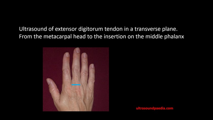
to examine the extensor apparatus of the finger:

  1. place the transducer transversely across the distal metacarpal just proximal to the knuckle. the extensor should be directly beneath the probe.
  2. slide the probe distally in transverse following the extensor digitorum to the base of the middle phalanx.
  3. rotate into longitudinal.
  4. confirm the ed insertion. 
  5. now move distally to confirm the extensor hood insertion to the distal phalanx.
  6. to assess each band of the extensor hood, keep the distal end of the probe on the insertion and rotate the proximal end very slightly to the radial, then ulnar aspects.
  7. dynamically assess the ed tendon sliding. at the level of the metacarpal head. importantly both in longitudinal and transverse (checking for tendon stability).

tips:

  • if you see a nice round ed tendon, it is probably abnormally thickened.

to examine the nail bed of the finger:

  1. place the transducer (++gel) longitudinally on the finger nail.
  2. ensure you can see the nail root proximally.
  3. slide sideways assessing the entire area.
  4. rotate into transverse and assess proximaly-distally.
  5. carefully check the underlying bony cortex for defects.
  6. check with colour doppler.

tips:

  • if you are uncertain, check neighbouring fingers.

normal appearance of the extensor tendons:

  1. transverse: echogenic and flat ovoid
  2. longitudinal: echogenic and fibrillar.
  3. size: <2mm
  4. no fluid should be visible.
  5. dynamics: the extensor tendon should glide freely and with stability at the level of the mcpj with finger flexion/extension.

normal appearance of the extensor hood:

  1. in the normal patient it cannot be seen independant of the extensor digitorum tendon.
  2. each band over the middle phallanx should be thin, parallel echogenic lines, shlightly hypoechoic centrally (with current technology)
  3. at the distal insertion it should be tapering, uniform, echogenic and fibrillar.

normal appearance of the nail bed:

  1. smooth underlying cortex
  2. hypoechoic nail matrix
  3. smooth, uniform parallel surfaces of the nail.
  4. the nail should taper evenly to a sharp nail root.
  5. uniform vascularity of the nail matrix with colour doppler.

once you identify the extensor apparatus of the finger:

  1. are they  fibrillar and homogeneous?
  2. do the gradually decrease in size distally?
  3. there should be no fluid visible.
  4. do they slide freely?
  5. does the ed stay in place at the metacarpal head during finger flexion (transverse plane is best). if the ed subluxes, there is a likely tear of the sagittal band of the extensor hood.
  6. are both bands of the distal extensor hood intact 9partial mallet finger)

once you identify the nail bed of the finger:

  1. is it uniform in thickness?
  2. is there any underlying bony irregularity/defect?
  3. is there a mass/collection?
  4. is there focal hyperaemia on colour doppler?

extensor digitorum

scan plane extensor hood insertion to the base of the distal phalanx.

ultrasound of the extensor digitorum tendon insertion onto the base of the middle phalanx.

extensor digitorum tendon in the transverse plane, at the level of the metacarpal head. 

assess dynamically with fist clenching. the tendon should only move slightly. if it slides sideways (subluxes), there is a likely extensor hood sagittal band tear.

extensor digitorum tendon in the transverse plane, at the level of the metacarpal head. 

assess dynamically with fist clenching. the tendon should only move slightly. if it slides sideways (subluxes), there is a likely extensor hood sagittal band tear.

extensor hood

extensor hood

  • the extensor hood overlies the extensor digitorum with fascial anchoring bands on each side.
  • at the level of the proximal interphalangeal joint it divides into 2 bands which rejoin for a common insertion on the base of the distal phalanx.

scan plane extensor hood insertion to the base of the distal phalanx.

ultrasound of the extensor hood insertion to the base of the distal phalanx.

note that the finger is slightly flexed to tighten the tendon.

nail bed

nail bed

assess uniformity of the underlying matrix and for a smooth, flat bone surface contour.

sac plane for the extensor digitorum insertion.

note the thick gel.

ultrasound of the nail bed is best viewed through a thick gel standoff.

ulna collateral ligament (ucl) of the thumb (mcpj)

ulnar collateral ligament of the thumb (ucl)

  • the ulnar collateral ligament of the 1st metacarpo-phalangeal joint.
  • a forced abduction/extension injury.
  • rupture is a skiier’s or gamekeeper’s thumb.
  • if the torn ligament folds under the adductor pollicis it is referred to as a ‘stenner lesion’.

dynamically assess

  • by gently flexing/extending the tip of the thumb, whilst scanning the immobilised ucl of the mcpj (this takes some practice).
  • the adductor should be seen as a very thin hypoechoic band sliding superficially over the ucl. if it is sliding over the bone at the joint, there is a stenner lesion.

the ulnar collateral ligament on the 1st metacarpo-phalangeal joint.

ultrasound of the ucl of the 1st mcpj : rupture is called a skiier’s or gamekeeper’s thumb. if the torn ligament folds under the adductor pollicis it is referred to as a ‘stenner lesion’.

scan protocol

role of ultrasound

to assess for:

  • muscular, tendinous and ligamentous damage (chronic and acute).
  • foreign bodies.
  • joint effusions.
  • soft tissue masses such as ganglia, lipomas.
  • classification of a mass eg solid, cystic, mixed.
  • post surgical complications eg abscess, oedema.
  • guidance of injection, aspiration or biopsy.
  • relationship of normal anatomy and pathology to each other.
  • some bony pathology.

limitations

  • open wounds
  • dressings
  • difficulty with probe contact due to curved /flexed surfaces.


patient preparation

  • nil

equipment setup

use of a high resolution probe (>12mhz)with a small footprint is essential when assessing superficial structures. careful scanning technique to avoid anisotropy (and possible misdiagnosis). beam steering or compounding can help to overcome anisotropy in linear structures such as tendons. good colour / power / doppler capabilities when assessing vessels or vascularity of a structure. be prepared to change frequency output of probe (or probes) to adequately assess both superficial and deeper structures.

common pathology

  • tendon tears
  • ligament tears
  • teno-synovitis
  • joint effusion
  • rheumatoid disease
  • foreign body
  • masses
  • fracture
  • ganglion
  • dupyetren’s
  • trigger finger
  • nail bed tumour

scanning technique

either:

  1. sit the patient on the side of the bed with a pillow on their lap to support their hand
  2. sit the patient on a chair on the opposite side of the bed with their hand resting on the bed.

joint effusions

for example images, click here to goto the hand & finger pathology page.

  • size
  • simple/complex
  • any synovial thickening
  • any vascularity on power doppler – normal is little or no discernable flow. hyperaemia = acute.

may need to compare with the other side.

tendon abnormalities

for example images, click here to go to the hand & finger pathology page.

look for hyperaemia, tendon sheath fluid (simple/complex) and tendon integrity/homogeneity

  • check for tendon thickening (compare with other side)
  • fluid in the tendon sheath
  • integrity of the tendon- any tear?
  • does the tendon slide freely when mobilised?

dupuytren’s contracture

what is it?

  • fibrosis of the palmer fascia forcing the flexion of the 4th/5th fingers.
  • gradual onset
  • m>f
  • often inherited.
  • generally affects 4th and 5th fingers.

scan in longitudinal from the base of the proximal phalanx down into the palm looking superficial to the flexor tendon

it will appear as a hypoechoic focal fusiform thickening of the palmar fascia at the metacarpal head level. not to be confused with trigger finger (see below).

trigger finger

for example images, click here to go to the hand & finger pathology page.

what is it?

tenosynovitis of a flexor digitorum tendon causing forced flexion of a finger.

initially in transverse, identify the flexor digitorum tendons at the metacarpal head level. follow the common tendon proximally to the carpal tunnel. then follow distally to the insertions: the flexor digitorum superficialis divides, with two slips inserting onto the side of the base of the middle phalanx. flexor digitorum profundus inserts onto the distal phalanx

finger pulleys

for example images, click here to go to the hand & finger pathology page.

what are they? bands of fibrous tissue holding the flexor tendon to the finger similar to runners on a fishing rod.

they are named according to their type-annular (around) or cruciform (cross), and numbered from proximal to distal. eg: a1 to a5 and c1 to c4.

scan longitudinally over the anterior surface of the finger. the pulleys may be seen as thin hypoechoic zones intimately overlying the flexor tendon sheath.

if ruptured, the tendon will no longer follow the bone and will instead “bowstring”.

game keepers thumb/ skiiers thumb

for example images, click here to goto the hand & finger pathology page.

what is it?

rupture of the ulnar collateral ligament of the thumb due to a sudden valgus force.
may occur after repeated stretching of the ligament.

the ligament usually tears at it’s distal end from the base of the proximal phalanx. if there is marked angulation of the phalanx, the flailing ligament may impinge under the adductor pollicis aponeurosis creating a ‘ stenner lesion ‘.

click here for detailed information.

foreign bodies

for example images, click here to goto the hand & finger pathology page.

ensure you approach the proposed site of the foreign body from different angles. some materials will be poorly reflective and almost invisible unless the beam is perpendicular to them. there will usually be a surrounding hypoechoic halo representing an inflammatory reaction

identify:

  • the plane of tissue it is in.
  • how close it is to the entry wound and to any blood vessels.
  • it may be helpful to mark the location and orientation of the foreign body on the skin to guide removal.

masses

95% of finger tumours are benign(ref: emedicine)

  • abscess
  • granuloma
  • ganglia
  • neuroma
  • fibroma
  • glomus tumour (nail bed tumour)

for non-specific palpable or visible masses see our superficial lumps page.

joint abnormalities

for example images, click here to goto the hand & finger pathology page.

  • gout: abnormal uric acid metabolism resulting in joint inflammation. may see tophaceous gout as a complex echogenic mass (tophus) in the soft
  • osteoarthritis: bony irreg at the bone ends with joint effusion. when acute the joint will be hyperaemic
  • rheumatoid arthritis: thickened synovium with a complex ‘thick’ joint effusion, pannus & associated bony irregularity

click here to read a great article on hand ultrasound.

basic hardcopy imaging

hard copy imaging should reflect the anatomy investigated.

  • document the normal anatomy. any pathology found in 2 planes, including measurements and any vascularity.