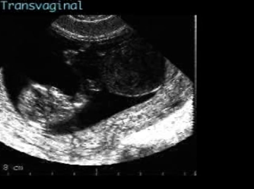
note the presence of a large, thin-walled cyst within the chorionic cavity, associated with the umbilical cord, distinct from the fetus and yolk sac, representing the allantoic cyst.
description
the allantois is a diverticulum of the yolk sac and develops at about the 16th day of gestation, joining with the fetal bladder prior to closure of the cloaca. the allantoic vessels develop into the umbilical vein and arteries. once the cloaca has divided, the allantois loses its connection with the hindgut although it remains connected to the bladder by the urachus which is eventually obliterated by about 6 weeks. remnants may be found in the umbilical cord but these usually disappear.
diagnosis
the appearance of a thin-walled cyst within the umbilical cord is usually diagnostic. colour doppler will indicate the avascular nature of the lesion. it is particularly important to confirm that the cyst is separate from the fetus, as this finding would imply the presence of a small omphalocele. in the absence of any other defects, the risk of associated anomalies is unlikely, although there may be an increased incidence of karyotypic abnormalities.
differential diagnosis
the most important lesion to exclude is a small omphalocele, and so care should be taken to ensure that the cyst is clearly separate from the fetus. angiomata of the umbilical vessels are occasionally seen, but these are vascular structures, clearly distinguishable using doppler. a thick cord, containing much wharton’s jelly, might present a similar picture. the bowel herniation associated with gastrochisis is usually distinct from the umbilical cord.
sonographic features
thin-walled cyst within the umbilicus at the fetal end
cyst is separate from the fetus
cyst does not usually increase in size and thus becomes smaller relative to the baby