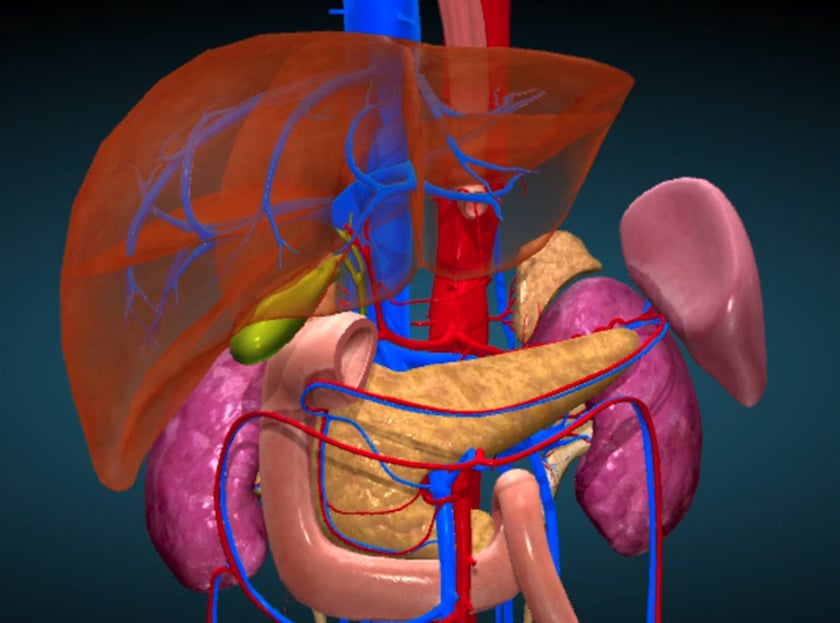
begin by doing a full survey sweep through the liver.
you will need the patient to take deep inspirations to fully visualise the superior borders of the liver.
look in transverse up and down the left lobe from a subcostal approach. look in transverse through the right lobe subcostally or intercostally.
roll the patient in a left lateral decubitus position for assessment of the rt lobe only after checking for fluid. bowel gas can overlie the liver in a subcostal approach, so getting the patient to distend their abdomen can help with visualisation. also looking intercostally between each rib space can ensure thorough visualisation.
look for:
- homogeneous v’s attenuative(normal v’s fatty)
- smooth v’s coarse echotexture
bmode image here
- size: to measure the size of the liver, use a sagittal approach in the mid clavicular line. measure from the diaphragm to the inferior border on bmode image. this can be very subjective. also look at the lower edge of liver in relation to the rt kidney.it should finish half way down the kidney. bmode image an enlarged liver will have rounded borders.
once you have thoroughly scanned though the liver, then start taking images.
normal liver measurements
- we need to be able to determine such conditions as hepatomegaly , splenomegaly, renal impairment and abdominal aortic aneurysm.
- it needs to be a consistent measurement to be able to compare sizes over time.
- the calipers need to be positioned in the same position between sonographers for accuracy.
- the upper border lies in the right midclavicular line at the 5th intercostal space.
(ref .diagnostic ultrasound 3rd edition volume 1 .rumack et al)
- most people have the lower border extending to the lower costal margin.
- if it is measured in the midhepatic line with a large field of view it should measure <16cm (ref : ultrasonic determination of hepatomegaly. j clin ultrasound 1981gosink bb)
from the posterior diaphragm to the lower anterior edge. however organ size increases with gender, age ,height, weight and body surface area.
- if the measurement is made from the anterior diaphragm to the lower edge of the liver in the midclavicular line it should be no >13cm (ref ultrasonography .an introduction to normal structure and functional anatomy:wb saunders; 1995.curry ra and tempkin bb.)
tip: be careful not to get confused with a riedel’s lobe as it can increase the measurement.