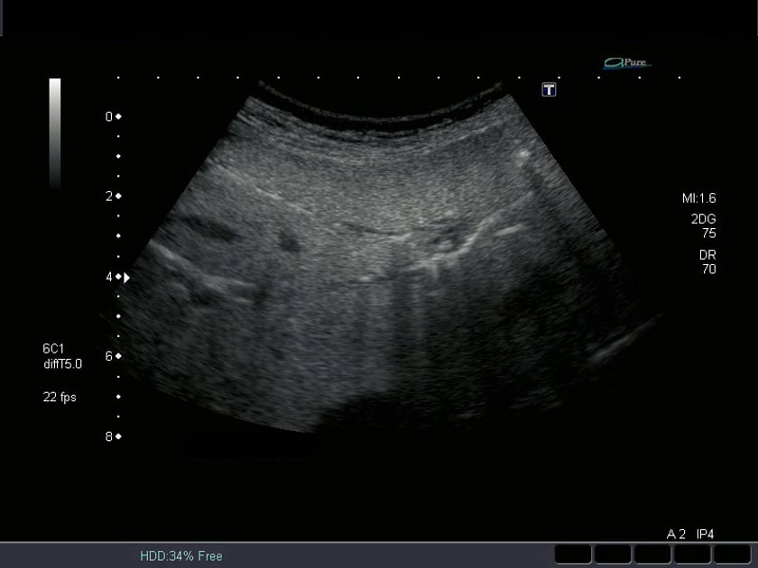
biliary tree pathology

cbd measurements

  • normal : <6mm (+1mm per decade over 60yrs)
  • post cholecystectomy : up to 10mm (but should gradually taper into the head of pancreas)

the common bile duct should never exceed 10mm in diameter.

ultrasound image- transverse view across porta hepatis.

ultrasound image- dilated cbd.

choledocholithiasis

choledocholithiasis is when there is at least one gallstone in the common bile duct.

about 15% of people with gallstones will develop stones in the common bile duct

risk factors include a history of gallstones. however, choledocholithiasis can occur in people who have had their gallbladder removed.

blockage and infection caused by stones in the biliary tract can be life threatening.

ultrasound image- stone impacted in the head of pancreas (hop)

ultrasound image- stones appear as echogenic rounded foci. there may not be any shadowing. sometimes the initial sign is that the bile duct is dilated. there may also be intrahepatic duct dilatation.

ultrasound image- a calculus is impacted in the distal cbd at the head of pancreas.

ultrasound image- mobile calculus:
by rolling the patient onto their side, you may be able to visualise the calculus rolling within the cbd, excluding impaction.this is demonstrated here where the same calculus from the image to the left has moved out of the distal cbd.

cholangitis

acute cholangitis is a bacterial infection generally related to biliary obstruction. the obstruction increases the risk of bacterial infestation in the cholestatic patient.
it can also occur post surgery/procedures that allow access of contaminants to the normally sterile bile. the most common such procedure being ercp.

on ultrasound, you may see thick walled ducts or pyogenic debris within the ducts.

see pictures & video.

primary sclerosing cholangitis is a chronic liver condition (thought to be auto-immune). it is characterised by fibrosis, cirrhosis and liver failure. there are no ultrasonically visible bile duct changes till later stage. early diagnosis is difficult with ultrasound.

this patient presented with extreme right upper quadrant pain with a history of gallstones. the bile ducts were inflamed. it is important to identify any stones in the ducts and report their position.

acknowledge: ms. alison deslandes.ams

ultrasound image- acute cholangitis
debris in the biliary tree related to biliary stasis and bacterial infection, secondary to obstruction.

caroli's disease

  • a rare, inherited disease.
  • involves non-obstrucive, saccular bile duct dilations.
  • usually a segmental distribution.
  • results in recurring cholangitis
  • poor long term prognosis with a high morbidity rate (secondary to complications of the recurring cholangitis +/- pancreatitis)
    on ultrasound:
  • early phase will see small, prominant and ectatic intrahepatic ducts which will develop into large cystic dilatations.

caroli’s disease
a family history and ruq symptoms, combined with the subtle ectasia of these dilated ducts leads to a likely diagnosis of early caroli’s disease.

cholangioma

cbd mass / cholangioma
a differential diagnosis is portal vein thrombosis, which can be readily excluded using colour doppler on the adjacent portal vein.

gas in the biliary tree

this is commonly seen folowing interventional procedures such as an ercp in which the sphyncter of oddi is disrupted to gain access to the cbd via the duodenum.
this results in bowel gas gaining access, and refluxing into the biliary tree.
occasionally there will be sufficient gas in the cbd to obscure adequate cbd visualisation with ultrasound.

ultrasound of gas in biliary tree.

intrahepatic duct dilatation

if uncertain, utilise doppler ultrasound to help differentiate vessels from ducts.

ultrasound image- intrahepatic duct dilatation


doudenal mass

ultrasound image- uncertain origin: invasive pancreatic v’s small bowel carcinoma.

ultrasound image- biliary dilatation secondary to the duodenal mass.

cbd stenting

ultrasound image- biliary dilatation secondary to the duodenal mass.

  • generally undertaken in cases of malignant obstruction
  • performed endoscopically via ercp (endoscopic retrograde cholangiopancreatography )
  • the stents are either plastic or metallic.
  • look for patency and position.

ultrasound image- pancreatic mass
a stent is visible within the mass in the head of te pancreas.
this is a palliative treatment to overcome the obstructive jaundice caused by either compression or invasion of the cbd by a pancreatic mass.

cbd stent
this ultrasound image shows extension of the pancreatic tumour into the lumen of the stent.
this re-obstructs the system and the patient may present with acute symptoms.

ultrasound of bile duct tumor stent.