breast pathology
breast cysts
simple v’s complex or complicated cysts
to be simple it must be:
- anechoic
- well circumscribed
- have posterior enhancement
- it’s height should not exceed it’s width.
ultrasound image- a simple breast cyst.
ultrasound image- a complex breast cyst:
sedimentary movement may be visible by scanning the patient erect.
fine needle aspiration and cytological assessment can confirm the diagnosis. at the least, a follow-up ultrasound should be performed.
ultrasound image- a simple cyst adjacent to a complex cyst, (confirmed by fna to be a haemorrhagic cyst).
colour doppler should be performed to assess for any internal vascularity.
ultrasound image- the material in this breast lesion is not in the dependent portion, therefore excluding mobile debris or fluid.
whilst it may represent a mural nodule, the neat linear edge suggests likely adherent haemorrhage.
a fine needle aspiration may still be warranted. or at least a 2 month follow-up scan.
ultrasound image- a complex, milk cyst.
fibroadenoma
- benign.
- well circumscribed solid ovoid mass with subtle posterior enhancement.
histological confirmation via a biopsy is still recommended. a core biopsy is preferable.
ultrasound image- well circumscribed solid ovoid mass with subtle posterior enhancement.
histological confirmation via a biopsy is still necessary. a core biopsy is preferable.
ultrasound image- fibroadenoma:
a phylloides tumour can have similar appearances and be indistinguishable from a fibroadenoma. if the mass is greater than 5cm or rapidly increasing in size a phylloides should be considered even if a fine needle biopsy has suggested a diagnosis of fibroadenoma.
ultrasound image- a core biopsy confirmed a very large fibroadenoma.
ultrasound of a large fibroadenoma in a 15 year old girl.
presented with asymmetry of breast size.
phylloides tumors (also called phyllodes)
- very similar to fibroadenomas in appearance.
- generally more rapidly growing.
- poorly differentiated by fine needle biopsy, so core biopsy is recommended.
ultrasound image- a phylloides tumour – confirmed by core biopsy.
note the similarities to a fibroadenoma.
ultrasound image- a large lobulated phylloides tumor.
papilloma
whist often benign, their malignant tendency generally leads to removal. multiple papillomas have been shown to carry a far greater risk than solitary.
(ref: ohuchi n, abe r, kasai m. possible cancerous change of intraductal papillomas of the breast. a 3-d reconstruction study of 25 cases. cancer. 1984;54:605.)
- they are fibrovascular growths within milk ducts behind the nipple.
- radiographic ductography has often been employed to confirm the diagnosis, however advancements in ductoscopy are proving to be of great benefit.
a document giving a proposed algorithm for management of suspected papilloma.
from: int semin surg oncol. 2006; 3: 1.
published online 2006 january 17. doi: 10.1186/1477-7800-3-1
ultrasound image- a papilloma in a markedly dilated duct.
ultrasound image-the same papilloma imaged in two planes.
ultrasound image-accessory nipple at the lateral areola.
ultrasound image-the nipple generally casts an acoustic shadow.
to overcome this use either a stand-off pad or thick gel.
ultrasound of the nipple with gel as a stand-off. a papilloma corresponding to the palbable nipple lump is visible.
ultrasound of a large papiloma in the breast nipple with and without colour doppler.
")
")
ultrasound of a papiloma in the breast nipple with and without colour doppler. gel has been used as a stand-off. this papilloma is in the tip of the nipple without causing any dusct dilatation.
breast carcinoma
common ultrasound appearance:
- poorly circumscribed, hypoechoic mass.
- height greater than width.
- posterior shadowing
you may also see: punctate, micro-calcifications, tethering of adjacent tissues or the mass crossing tissure boundaries.
elastography is also an emerging technique in assisting suspicion levels.
types of breast cancer
**** breast cancer grading and specific differentiation must involve a series of investigations and not be based on ultrasound alone.
carcinoma in-situ
indicates that the cancer is still contained entirely with the tissue of origin and not penetrated tissue boundaries (a histological diagnosis)
- ductal carcinoma in-situ (dcis) – the cancer originated within breast milk ductal epithelium and is still contained by the ductal walls.
- lobular carcinoma in-situ (lcis)–
infiltrating carcinoma
as the name suggests, the cancer has crossed multiple tussue boundaries, and is no longer contained in the tissue of origin.
- infiltrating ductal carcinoma (idc)
- infiltrating lobular carcinoma (ilc)
there are several grades using the tnm grading
medullary carcinoma
- has better defined margins so has a better prognosis than ductal or lobular. only accounts for approximately 5% of breast cancers.
colloid (or mucinous) carcinoma
- rarer again. arises from mucous secreting cells. also a better prognosis.
tubular carcinoma
- is a form of ductal carcinoma with tubular cells visible on histo-cytology. with increasing early (sub-clinical) screening, tubular carcinomas are being detected with increased frequency.
paget’s disease of the breast
- greater than 97% of patients with pagets diseaes of the nipple have an underlying breast cancer (ref breastcancer.org)
- accordingly, accurate diagnosis is important.
- clinically, the patient may have an eczema-like rash around the nipple/areola and nipple discharge. there may be itching/tingling or hypersensitivity of the nipple.
- as many pagets related breast cancers begin in the ducts behind the nipple, ensure this area is scanned thoroughly with high resolution equipment.
- mammography and breast mri are appropriate investigations in these patients.
inflammatory breast cancer
- rare but aggressive
- the cancer blocks the lymphatic drainage of the cutaneous tissues.
- unexplained onset of reddened, swollen, firm breast in the abscence of infection.
ultrasound image – the posterior shadowing and irregular outline raises the level of suspicion from fibroadenoma to possible carcinoma.
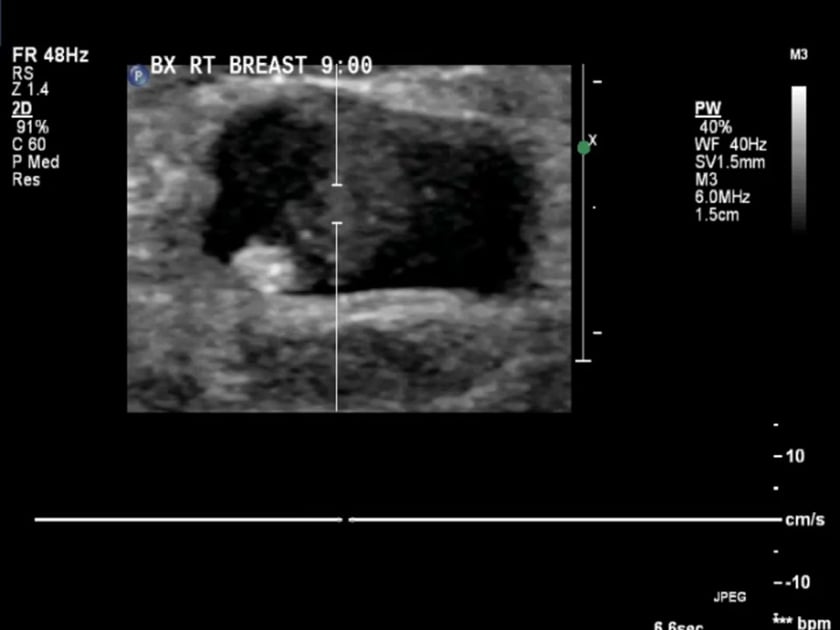
ultrasound biopsy image- the core biopsy needle is seen to transverse this complex lesion.
ductal carcinoma
dcis is by far more common than lcis, and more importantly, it should be distinguished as a clearly malignant lesion. ductal epithelial cells undergo malignant transformation and proliferate intraluminally. eventually, the cells outstrip their blood supply and become necrotic centrally. this debris can calcify and be detected mammographically. moreover, the lesions also may be palpable clinically. five pathologic subtypes have been identified: comedo, papillary, micropapillary, solid, and cribriform. most lesions represent a combination of at least two of these subtypes.
the presence of comedo necrosis is an independent risk factor for subsequent ipsilateral breast cancer (nsabp-b17).
ref: http://emedicine.medscape.com
ultrasound image – this focal ductal carcinoma is easily seen.
the red arrows indicate the microcalcifications.
ultrasound image- an invasive ductal carcinoma of the breast.
power doppler ultrasound demonstrating subtle flow. this confirms the solid nature of the lesion (versus complicated cyst, abscess or haematoma), but doesn’t help differentiate between cancer types.
infiltrating lobular carcinoma
infiltrating lobular carcinoma has a much lower incidence and comprises less than 15% of invasive breast cancer. it is characterized histologically by the indian file arrangement of small tumor cells. like ductal carcinoma, these typically metastasize to axillary lymph nodes first. however, it also has a tendency to be more multifocal. despite this, the prognosis is comparable to that of ductal carcinoma.
ref: http://emedicine.medscape.com
ultrasound image- infiltrating lobular carcinoma.
ultrasound image- lobular breast carcinoma. longitudinal.
ultrasound image – an invasive lobular carcinoma invading the latissimus dorsi. this particular image demonstrates the use of harmonics to better define the irregular and spiculated margins.
image courtesy of callum linehan.
ultrasound image – longitudinal to the latissimus dorsi. this demonstrates the invasive nature of a metastatic lobular carcinoma, seen infiltrating the latissimus dorsi and subcutaneous tissue.
image courtesy of callum linehan.
the deceiving false positive
this ultrasound was undertaken and was followed with a biopsy the next day. on imaging it was highly suspicious for an carcinoma. it was surprisingly found to a fibroadenoma.
this ultrasound image does not follow the rule of fibroadenomas being wider than deeper in size. it also has an irregular border.
ultrasound image- it has internal low resistance internal flow.
thankfully it was a benign fibroadenoma.
gynaecomastia
- is the abnormal enlargement of rudimentary male breast tissue.
- can be idiopathic, related to hormonal fluctuations(puberty), steroid abuse or associated with hormonal treatments such as prostate cancer therapies.
- may present as a retroareolar lump with or without pain.
- on ultrasound it will be hypoechoic with spiculations radiating away from the nipple.
ultrasound image- male breast gynaecomastia – mild.
a tender (often) lump deep to the nipple with spiculations radiating away from the nipple.
can be unilateral or bilateral.
ultrasound image- male breast gynaecomastia-acute. mildly increased vascularity can be seen.
can be idiopathic, related to steroid abuse or associated with hormonal treatments such as prostate cancer therapies.
breast implants
- should be scanned with the patient positioned as normal.
- treat the scan as a 2-fold examination:
- the breast tissue.
- the deeper implant. this may require lower frequency or a curved probe to investigate.
the implant should be anechoic with well defined margins. folds are commonly seen in the implant surface.
also, small traces of simple fluid will be seen overlying the implant but is contained by the overlying fibrous capsule that contains the implant. this fluid is routinely seen within the implant folds (see image below).
most saline implants will have a small valve visible (see image below)
the normal appearance of a fold in an implant with normal physiological amount of capsular free fluid. the mammogram below shows the typical appearance of folds.
ultrasound image- a normal finding of an implant ‘valve’. also visible in the mammogram below.
the typical appearance of folds in a breast implant.
ultrasound image- most often there is a rupture only of the elastomer biluminal shell of the implant. the ruptured material is then contained by the fibrous capsule generated by the body.
ultrasound image- silicone extruded outside the fibrous capsule. this can be seen as an island of poorly defined echogenic material ,distant to the implant, casting a ‘dirty’ shadow .
ultrasound image- this implant has a large lateral rupture with silicon extruded out (green). this tends to alter the internal silicone also (orange).
ultrasound image- the silicone has migrated into the lateral chest wall lymph node (green). the silicone has the pathognomonic echogenic ‘dirty shadow (yellow).
ultrasound image- a large siliconoma.
this is a palpable lump of silcone. the cystic space within the silicon is a commonly seen variant.
ultrasound image- silicon has made its way into the intercostal space.
ultrasound image- an acutely ruptured implant.
there is discontinuity of the implant capsule but most of the silicone is contained by the fibrous capsule created by the body.
ultrasound image- the same impalnt rupture showing the collapsed implant capsule (curved parallel lines).
ultrasound image- old, degenerated, intact silicone implant.
always increase the gain on your ultrasound to examine the ‘quality’ of the implant.
ultrasound image- an extracapsular ruptured silicone implant.
the diffuse silicone ‘cloud’ with no visible implant capsule.
ultrasound image- the left silicone implant had degenerated and burst. here it is still contained by the fibrous capsule.
collections and infections
seroma
- a post-operative seroma is not an uncommon finding in the post mastectomy patient.
- they can be very painful and rapidly increase in size.
- therapeutic ultrasound guided fine-needle aspiration can be performed as required to relieve symptoms.
ultrasound image- a post-operative seroma is not an uncommon complication post mastectomy.
panoramic view of a breast seroma.
haematoma
- may occur following trauma, biopsy or surgery.
- seatbelt injuries cause substantial cutaneous bruising. this usually appears on ultrasound as diffusely increased echogenicity of the underlying breast tissue, perhaps with some small hypoechoic collections.
panoramic ultrasound of an extensive breast haematoma.
a large, expansive breast haematoma adjacent to a breast implant.
abscess
- acutely tender lump or swelling with erythema.
- may be secondary to lactating mastitis.
ultrasound image- a breast abscess. note the thickened wall and complex contents. if any clinical doubt, aspiration checking cytology and microbiology (culture and sensitivity should be performed).
mondor cord
this condition is sclerosing thrombophlebitis of the subcutaneous veins of the anterior chest wall. it is associated with patients who have had breast surgery such as lumpectomy for breast cancer or breast augmentation.
- the patient will present with a visible thin tight ridge. inframammary is mosy common. also seen in the axilla.
- not usually painful but can feel tight/tethering and can be worrying to the patient.
- usually resolve and do not require intervention however if recalcitrant, may require intervention.
- despite their obvious visual clinical presentation, they can be difficult to visulise on the ultrasound scan because they are often very thin, very superficial and isoechoic. high frequency linear probe, using a standoff or alot of gel will assist.
- important to ensure there is no local deep venous extension
ultrasound image- it is a small cord like structure/vein which does not have any vascularity.
ultrasound image- it can be very small and extremely difficult to do a panoramic view . a thorough history should be taken from the patient.
breast infection / mastitis / abscess.
ultrasound image- a breast abscess. note the thickened wall and complex contents. if any clinical doubt, aspiration checking cytology and microbiology (culture and sensitivity should be performed).