the important feature is the similarity between the abnormal elements within the fetus and in the external mass.
description
a fetus-in-fetu is an encapsulated, pedunculated vertebrate tumor. it represents a malformed, monochorionic diamniotic parasitic twin included in a host (or autosite) twin. characteristically the fetus-in-fetu complex will be composed of a fibrous membrane (equivalent to the chorioamniotic complex) that contains some fluid (equivalent to the amniotic fluid) and a fetus suspended by a cord or pedicle. the presence of rudimentary spinal architecture is used to differentiate a fetus-in-fetu from a teratoma, since teratomas are not supposed to develop through the primitive streak stage (12-15 days). this aspect has been considered too stringent by many authors who regard rudimentary body architecture (metameric segmentation, craniocaudal and lateral differentiation, body coelom) as an equivalent criterion. although teratomas can achieve striking degrees of differentiation by the inductive effect of adjacent tissues on one another, they do not present the criteria mentioned above. they also occur predominantly in the lower abdomen, not the upper retroperitoneum. teratomas also have a definite malignant potential, a feature that has not been reported in fetus-in-fetu. however, the coexistence of a fetus-in-fetu and a teratoma as well as the occurrence of a teratoma 14 years after removal of a twin fetus-in-fetu has been reported, supporting the older hypothesis of a continuum between twin and teratoma. cases of sacrococcygeal fetus-in-fetu should probably be regarded and treated as teratoma, because of the high incidence of teratoma in this region. ectopic testicles have a higher incidence of germ cell tumors, and the differentiation between fetus-in-fetu and teratoma is particularly important. some have argued that fetus-in-fetu should be considered as a teratoma since they do not evolve into lithopedion like fetuses of abdominocyesis. that argument is probably not valid since in abdominocyesis the antigen complements of the host and fetus are different, contrasting with fetus-in-fetu.
diagnosis
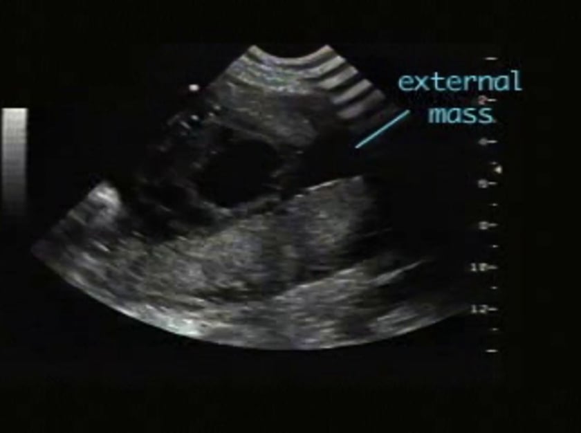
the few cases detected prenatally all presented as a complex mass. the general appearance is of a well-delineated capsule, with an echogenic mass suspended in fluid or partially surrounded by fluid. occasionally, the diagnosis may be suggested by the recognition of a rudimentary spine.
differential diagnosis
prenatally, the main differential diagnosis is with teratoma. by differentiation and induction, they can achieve striking organization, with examples of several organs being well formed. however, teratomas do not have vertebral segmentation, craniocaudal and lateral differentiation, body coelom or systemic organogenesis. thus the presence of a mass with a spinal organization and surrounded by fluid suggests the correct diagnosis.
sonographic features
complex solid/cystic mass within fetus
spinal component may be evident
associated syndromes
none
references
sada i, shiratori h, nakamura y antenatal diagnosis of fetus in fetu asia oceania j obstet gynaecol 12:353-356