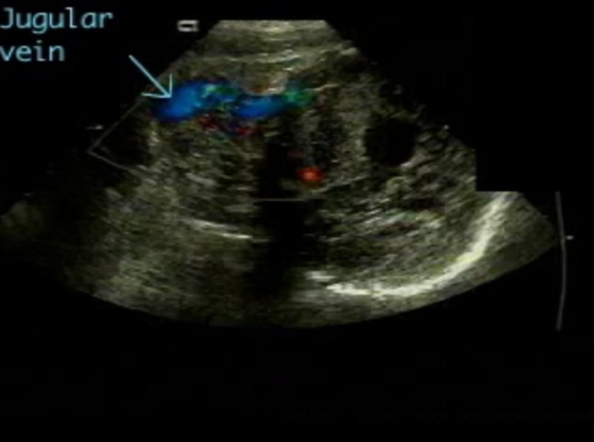
arteriovenous malformations
description
the two internal cerebral veins unite just posterior to the pineal gland to form the great vein of galen, which then courses posteriorly to empty into the straight sinus. aneurysms of this vein are actually end-to-end anastomoses of a major artery to a vein without the usual intervening capillaries. this abnormality, sometimes erroneously called an aneurysm, is a gross dilatation of the vein of galen, being fed directly by a large anomalous vessel or vessels arising from the carotid or basilar circulation.
diagnosis
it appears that fetal arteriovenous malformations may develop in or enlarge to a detectable size over a short period of time, usually in the third trimester. to date, no vein of galen aneurysm has been reported prior to 30 weeks gestation. the sonographic findings in this disorder are typical. in the transthalamic plane, a round or ovoid hypoechoic mass is seen posterior to the third ventricle. a tubular extension of the mass, representing a dilated straight sinus, can be traced posteriorly to the enlarged torcular of herophilus (confluence of dural sinuses) which has a triangular shape. the combination of a dilated vein of galen, straight sinus and torcular results in a key-hole shaped sonolucency. doppler examination of the abnormality may show arterial and/or venous flow, depending on the area sampled. the posterior cerebral artery and its branches are the most commonly involved feeders to this malformation, resulting in turbulence at the confluence of arterial and venous streams. occasionally this may be seen without resorting to doppler scanning. ventriculomegaly may be observed secondary to aqueductal compression. the disorder is associated with a poor outcome, but the infant’s chance of survival can be maximized by planning delivery at a tertiary care centre appropriately equipped to manage the affected newborn. heart failure, rather than hydrocephalus, is the major finding in symptomatic newborns. the antenatal appearance of the heart is variable, but generalized cardiomegaly or right ventricular enlargement is the rule. coarctation of the aorta and transposition of the great vessels occur with increased frequency in infants with this abnormality.
differential diagnosis
blood flow within the lesion differentiates it from other midline cystic structures, which include: cysts of the cavum septi pellucidi dilated third ventricle arachnoid cysts intracranial tumours intracranial haemorrhage if doppler studies demonstrate flow, an arteriovenous character (low resistance index, pronounced end-diastolic flow) will differentiate it from a true intracranial aneurysm.
sonographic features
inter-hemispheric midline tubular cystic structure, superior and posterior to the thalami.
doppler study shows blood flow in ‘cyst’ with venous component cardiac malformations may coexist.
associated syndromes
- none
references
- oberhoffer r, grab d, terinde r, lang d echocardiographic and sonographic clues for the prenatal diagnosis of cerebral arteriovenous malformations j matern fetal invest 4; 51-54
- ballester m-j, raga f, serra-serra v, bonilla-musoles f early prenatal diagnosis of an ominous aneurysm of the vein of galen by color doppler ultrasound sonography acta obstet gynecol scand 73: 592-595
- rodesch g, hui f, alvarez h, tanaka a, lasjaunias p prognosis of antenatally diagnosed vein of galen aneurysmal malformations childís nerv syst 10: 79-83
- dan u, shalev e, greif m, weiner e prenatal diagnosis of fetal brain arteriovenous malformation: the use of color doppler imaging j clin ultrasound 20:149-151
- koven mb, cohen hl, goldman ma fetal intracranial avm presenting as enlarged cardiac chamber j ultrasound med 12: 177
- comstock ch, kirk js arteriovenous malformations: locations and evolution in the fetal brain j ultrasound med 10: 361-365
- strauss s, weinraub z, goldberg m prenatal diagnosis of vein of galen arteriovenous malformation by duplex sonography j perinat med 19: 227-230
- suma v, marini a, saia os, rigobello l vein of galen aneurysm the fetus 1: 7478.1
- funk kc, siegel mj sonography of congenital midline brain malformations radiographics 8: 11-25
- sepulveda w, platt cc, fisk nm prenatal diagnosis of cerebral arteriovenous malformation using color doppler ultrasonography: case report and review of the literature ultrasound obstet. gynecol. 6: 282-286
- doren m, tercanli s, holzgreve w prenatal sonographic diagnosis of a vein of galen aneurysm: relevance of associated malformation for timing and mode of delivery ultrasound obstet. gynecol. 6: 287-289