polyhydramnios
description
polyhydramnios is defined as a volume greater than 2000 ml at term, a maximal vertical pocket of 8 ml or greater, or an afi above the 95th percentile. the reported prevalence of polyhydramnios ranges between 0.4 and 3.5 percent, with the frequency of diagnosed polyhydramnios at a given institution dependent upon method of ascertainment and proportion of high-risk patients seen. normally amniotic fluid volume increases steadily from about 250 ml at 16 weeks gestation to 800 ml at 28 weeks, 1000 ml at 34 weeks, and declines to approximately 800 ml at term. the actual volume is determined by a steady state between input: fetal urine (400-1200 ml/da), alveolar exudate (600-900 ml/da) and outflow: fetal swallowing (200-450 ml/da), and reabsorption through chorionic membranes (80 ml/da). input is affected by maternal blood flow to the placenta and the capacity for fetal maternal exchange at the intervillous space, modulated by maternal serum osmolality and maternal intravascular volume. acute polyhydramnios is uncommon (approximately 2-3% of cases of polyhydramnios) and is characterised by a rapid increase in amniotic fluid volume. the acute form occurs at an average gestational age of approximately 23-24 weeks, while the chronic form frequently occurs later. the acute form is often seen in association with monozygotic twin gestations and has been reported to complicate twin pregnancy approximately 2% of the time. in these cases fetal polyuria of the hyperfused twin is considered to be the cause. polyhydramnios is also associated with maternal diabetes, maternal syphilis, fetal macrosomia, rh isoimmunization, non-immune hydrops, fetalis, multiple gestations, and lesions of the umbilical cord and placenta. damato et al analyzed 105 second and third trimester pregnancies with amniotic fluid pocket 7.8 cm. overall, 63% of the pregnancies with polyhydramnios had anomalies. the most common anomaly in singleton pregnancies were gastrointestinal abnormalities. the most common anomalies in twin pregnancies with polyhydramnios was twin-to-twin transfusion syndrome, seen in 63% of affected twin pregnancies. in 29 patients who underwent amniocentesis, 24% were chromosomally abnormal (trisomy 21, 18, 13). all babies with abnormal karyotypes had identifiable anomalies on ultrasound. potential mechanisms for polyhydramnios related to fetal malformations include: 1) impaired fetal swallowing or gastrointestinal reabsorption including obstructive neck masses and neurologic impairment. 2) abnormal fetal renal function (associated with 25% of upj obstructions in one series). 3) transudation across an exposed membranous lesion (e.g. cns anomalies etc.). 4) mass lesions associated with fetal congestive heart failure (hydrops, sacrococcygeal teratoma etc.) polyhydramnios is associated with an increased perinatal morbidity and mortality secondary to prolapsed cord, abnormal presentations, premature rupture of membranes (prom), premature labour, placental abruption, post partum haemorrhage and dysfunctional labour.
diagnosis
diagnosis is made either by subjective assessment by an experienced examiner, or by utilising semiquantitative methods such as an afi above the 95th percentile or an afi above 24 cms. the single vertical pocket method can also be used and an 8 cm 12 cm pocket is considered mild polyhydramnios; 12 cm-16 cm, moderate; and 16 cm or greater, severe polyhydramnios. attempts to reduce amniotic fluid volume (e.g. prostaglandin synthetase inhibitors) may be monitored by following serial afis. assessment of the fetus for gross malformations is essential in determining the aetiology of polyhydramnios.
differential diagnosis
a maternal ovarian cyst or very full maternal bladder can be easily identified in an extrauterine location.
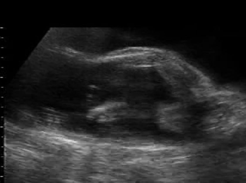
sonographic features
increased amniotic fluid volume (for gestational age)
assessment can be subjective (experienced examiner)
amniotic fluid index (afi) and the single vertical pocket methods are currently used to quantify amniotic volume especially in the third trimester
for single vertical pocket 8 cm-12 cm is considered mild; 12 cm-16 cm, moderate; and greater than or equal to 16 cm, severe polyhydramnios
for afi percentile values are available from 16 to 42 weeks and an afi above the 95th percentile at these gestational ages indicates polyhydramnios
associated syndromes
- (ccam) congenital cystic adenomatoid malformation
- achondroplasia
- arthrogryposis multiplex
- bartter
- beckwith-wiedemann
- camptomelic dwarfism
- congenital cytomegalovirus infection
- congenital myotonic dystrophy
- congenital toxoplasmosis infection
- erythroblastosis fetalis (maternal rh sensitization)
- fetal goitre/teratoma
- hydrolethalus
- jeune asphyxiating thoracic dystrophy
- klippel-feil
- maternal diabetes
- maternal myasthenia gravis
- nager acrofacial dysostosis
- osteogenesis imperfecta (ekman lobstein type)
- pena shokeir
- pentasomy x
- sacrococcygeal teratoma
- thanatophoric dwarfism
- trap sequence (twin reversed arterial perfusion)
- treacher collins
- trisomy 13
- trisomy 18
- trisomy 21
- twin-twin transfusion
references
chervenak fa, isaacson gc, campbell s in: ultrasound in obstetrics and 足球世界杯赛程2022赛程表
vol i little, brown, & co: boston, p555-563
chervenak fa, isaacson gc, campbell s in: ultrasound in obstetrics and 足球世界杯赛程2022赛程表
vol i little, brown, & co: boston, p1063-1081
chervenak fa, isaacson gc, campbell s in: ultrasound in obstetrics and 足球世界杯赛程2022赛程表
vol i little, brown, & co: boston, p565-568
nyberg d, mahony b, pretorius d in: diagnostic ultrasound of fetal anomalies vol ii mosby year book: st. louis, p50-66
damato n, filly ra, goldstein rb, callen pw, goldberg j, golbus m frequency of fetal anomalies in sonographically detected polyhydramnios ob/gyn survey 48:453-54
rachelson b, wagner j, shmoyss the clinical significance of resolving polyhydramnios ultrasound obstet gynecol 2:321