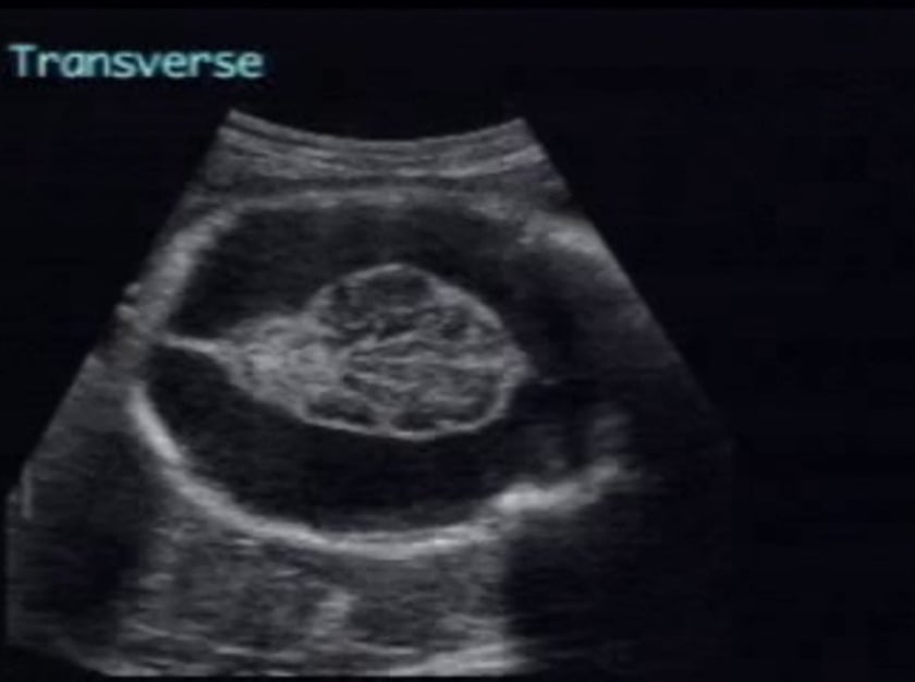
porencephaly
description
porencephaly implies replacement of cerebral cortex by cystic cavities or clefts (schizencephaly). these may represent extensions of the subarachnoid space in regions where there has been maldevelopment of cerebral tissue or where traumatic or ischemic episodes have taken place. hydranencephaly may therefore be considered an extreme form of porencephaly.
diagnosis
intraparenchymal cysts are readily seen on ultrasound. single or multiple cysts are noted, often communicating with the lateral ventricles or subarachnoid space. in schizencephaly, bilateral clefts are typically seen; though because of near calvarial reverberation, it is possible to miss a near side lesion and make a diagnosis of isolated unilateral porencephaly. due to ventricular communication there may be consequent ventriculomegaly (hydrocephaly ex vacuo) with marked asymmetric dilatation of the lateral ventricles and occasional displacement of the midline echo. the cysts and clefts exert no mass effect on adjacent structures. serial scans are recommended to monitor progression. the finding of a midline parietal scalp anomaly (i.e. cephalocele) in association with hydrocephalus and a prominent midline intracranial cyst is a form of congenital midline porencephaly.
differential diagnosis
with larger porencephalic cysts: symmetrical enlargement of the lateral ventricles, together with identification of falx and cerebral cortex will suggest obstructive hydrocephaly. the absence of even a rim of cortex, together with the characteristic bulging of thalami and midbrain into the cystic cavity will imply hydranencephaly. associated facial defects in the presence of a monoventricle will be diagnostic of alobar holoprosencephaly. with smaller porencephalic cysts: arachnoid cysts may be distinguished by the mass effect they exert on adjacent structures, and they do not communicate with the lateral ventricles. the interhemispheric cyst seen in agenesis of the corpus callosum, the dilated third ventricle, is characterised by its midline site and the typical appearance of the lateral ventricles in the transverse plane. cysts of the cava septi pellucidi et vergae are to be found in a central midline location, not typical for a porencephalic cyst.
sonographic features
echolucent cysts or clefts replacing brain parenchyma, often communicating with the lateral ventricles or subarachnoid space.
single or multiple defects seen.
may cause hydrocephaly with marked asymmetric dilatation of lateral ventricles.
no mass effect.
associated syndromes
- chorionic villus sampling
- encephalocutaneous lipomatosis
- epidermal nevus
- generalized fibromatosis-cutis marmorata
- glass
- l’hermitte
- oculo-cerebro-cutaneous
- pycnodysostosis
references
suchet ib schizencephaly: antenatal and postnatal assessment with colour-flow doppler imaging can assoc radiol j 45: 193-200
kormaniski ca, cyr dr, mack la, weinberger e prenatal diagnosis of schizencephaly j ultrasound med 9: 305-307
tom¹ p, lucigrai g, ravegnani m, cariati m, magnano g, lituania m hydrocephalus and porencephaly: prenatal diagnosis by ultrasonography and mr imaging j comput assist tomogr 14: 843-845
patten rm, mack la, nyberg da, filly ra twin embolisation syndrome: prenatal sonographic detection and significance radiology 173: 685-689
lituania m, passamonti u, cordone ms, magnano gm, toma p schizencephaly: prenatal diagnosis by computed sonography and magnetic resonance imaging prenat diagn 9: 649-655
vintzileos am, hovick tj, escoto dt, weinbaum pj, campbell wa, nochimson dj congenital midline porencephaly: prenatal sonographic findings and review of the literature am j perinatol 4: 125-128
klingensmith wc, cioffi-ragan dt schizencephaly: diagnosis and progression in utero radiology 159: 617-618